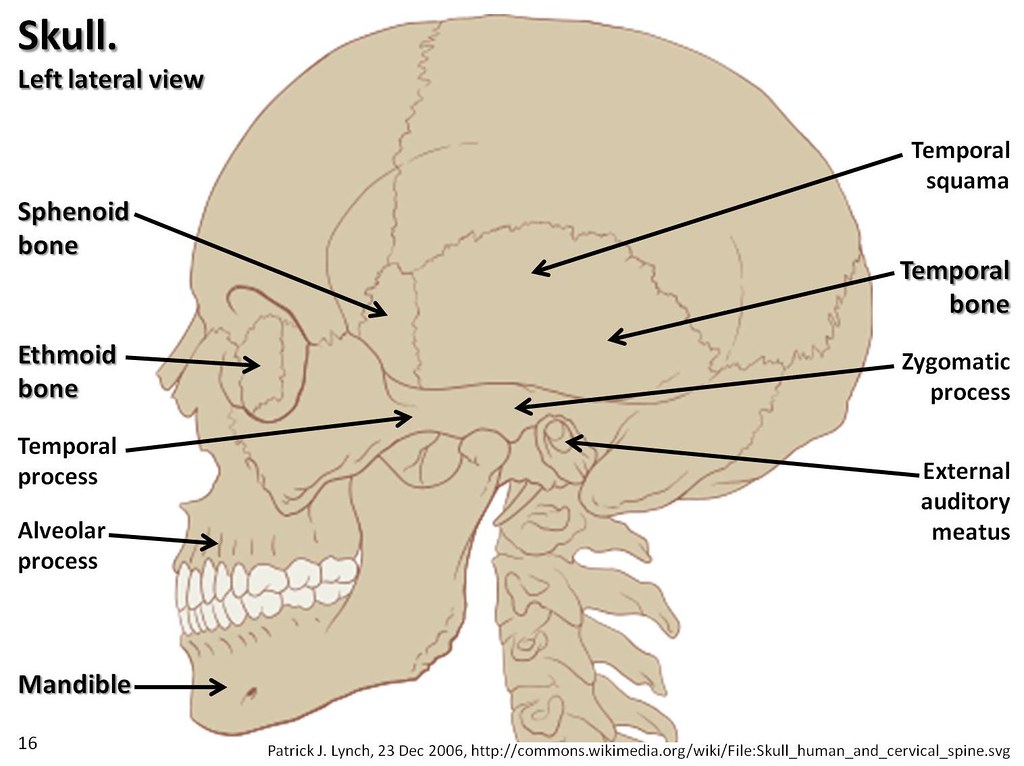

This bony prominence can vary from narrow and pointed to broad and flat.Passing through the superior orbital fissure from the middle cranial fossa are 1) the superior and inferior branches of the oculomotor nerve, 2) the trochlear nerve, 3) the abducent nerve, 4) the lacrimal, frontal, and nasociliary branches of the ophthalmic nerve, 5) the superior ophthalmic vein 6) A recurrent meningeal artery if present6) The squamous part of the temporal bone:It contains the following foraminaand landmarks1) Foramen rotundum(located in the greater wing of the sphenoid) transmits maxillary nerve. The junction of the upper and lower segments of the lateral edge (from the orbital side) is the site of a bony prominence that serves as the site of attachment of the lateral edge of the annular tendon, from which the four rectus muscles arise. The fissure is not oriented in a strictly coronal plane, but is directed forward so that the lateral apex is slightly forward of the medial margin. The fissure slopes gently downward from its lateral to medial border. From the orbital side the frontal bone forms a small portion of the lateral apical margin of the fissure, because the greater and lesser wings approach, but do not meet at the narrow lateral apex.It has a somewhat triangular shape, having a wide base medially on the sphenoid body and a narrow apex situated laterally between the lesser and greater wings. The petrous ridge complete the circle between lingual process and posterior clinod to form end of carotid canal.5) Superior orbital fissure: The superior orbital fissure is located between the roof of orbit (lesser wing of the sphenoid bone) and lateral wall of orbit (greater wing of the sphenoid bone ) on the lateral side of the optic canal. Below the lingula & the carotid groove found the petrygoid canal. 4) The lingual of the sphenoid body: is the most medial part of the edge of greater wing that prolonged backward over the carotid groove on the side of body. 2) The greater wing of sphenoid: The great wings, or ali-sphenoids, are two strong processes of bone, which arise from the sides of the body, and are curved upward, lateralward, and backward.3) the sphenoidal spine: the posterior part of each projects as a triangular process which fits into the angle between the squama and the petrous portion of the temporal and presents at its apex a downwardly directed process. Lesser wings of sphenoid bone terminate in a curved knife-edge, the inferior aspect of which contains the sphenoparietal sinus. The optic canal transmits the optic nerve and the ophthalmic artery and sympathetic plexus around it.

The lateral part is formed by 1) The lesser wing of the sphenoid bone: which blends medially into the anterior clinoid process.The lesser wing is connected to the body of the sphenoid bone by two roots, the upper (or anterior) thin and flatan root, which forms the roof of the optic canal, and by a lower (or posterior) thick and triangular root, also called the optic strut, which forms the floor of the optic canal and separates the optic canal from the superior orbital fissure.Pathology in Central skull base in Head and Neck Imaging. (2003): Central Skull Base: Embryology Anatomy, and Posterior condylar canal The condylar fossa


 0 kommentar(er)
